Diabetic Retinopathy
What is diabetic eye disease?
 Diabetic eye disease refers to a group of eye problems that people with diabetes may face as a complication of diabetes. All can cause severe vision loss or even blindness.
Diabetic eye disease may include:
- Diabetic retinopathy—damage to the blood vessels in the retina.
- Cataract—a clouding of the eye’s lens. Cataracts develop at an earlier age in people with diabetes.
- Glaucoma—increase in fluid pressure inside the eye that leads to optic nerve damage and loss of vision. A person with diabetes is nearly twice as likely to get glaucoma as other adults.
What is diabetic retinopathy?
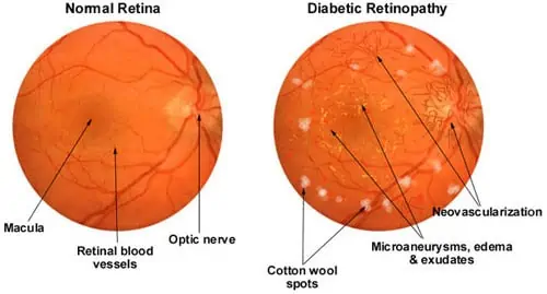
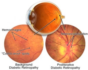
Diabetic retinopathy is the most common diabetic eye disease and can seriously affect vision and in some cases may cause blindness. In some people with diabetic retinopathy, blood vessels may swell and leak fluid. In other people, abnormal new blood vessels grow on the surface of the retina. The retina is the light-sensitive tissue at the back of the eye.
What are the symptoms?
- Often there are no symptoms in the early stages of the disease, nor is there any pain. If you have diabetes (whether Type 1 or Type 2), you should have a dilated eye examination at least once a year by an Ophthalmologist. Don’t wait for symptoms.
- Blurred vision may occur when the macula—the part of the retina that provides sharp central vision—swells from leaking fluid. This condition is called macular edema.
- If new blood vessels grow on the surface of the retina, they can bleed into the eye and block vision.
Normal Vision and the same scene viewed by a person with diabetic retinopathy.


Who is at risk?
- Poorly managed diabetes or high blood sugar levels
- High blood pressure and also have kidney disease
Diagnosis
Diabetic retinopathy is detected during an eye examination that includes:
- Visual acuity test: This test uses an eye chart to measure how well you can see at various distances
- Tonometry: An instrument that is used to measure the pressure inside the eye.
- Pupil dilation: Eye drops are put into your eyes to dilate the pupils. This allows the doctor to see more of the retina and look for signs of diabetic retinopathy. After the examination, close-up vision may remain blurred for several hours.
- Ophthalmoscopy: This is an examination of the retina in which the doctor looks through a slit lamp biomicroscope with a special magnifying lens that provides a narrow view of the retina.
The doctor will look at the retina for early signs of the disease, such as:
- Leaking blood vessels
- retinal swelling, such as macular oedema
- pale, fatty deposits on the retina (exudates) – signs of leaking blood vessels
- damaged nerve tissue (neuropathy)
- any changes in the blood vessels.
Should the doctor suspect macular oedema, he or she may Optical Coherence Tomography (OCT) and sometimes perform fluorescein angiography.
- Optical coherence tomography (OCT): Optical Coherence Tomography (OCT) is a non-invasive diagnostic imaging technique that uses light to produce very high-resolution cross-sectional images of the tissue layers within the retina which can be used to measure the thickness of the retina and to resolve its major layers, allowing the observation of swelling and/or leakage.
- Fundus Fluorescein angiography (FFA): This is an imaging technique which relies on the circulation of Fluorescein dye in the eye vasculature. It involves an injection of Sodium Fluorescein into the systemic circulation and obtaining a series of photographs. The fluorescein dye also reappears in the patient urine, causing a yellow-green appearance.
Risks of FFA
There is a slight chance of infection any time the skin is broken. Rarely, a person is hypersensitive to the dye and may experience:
- Dizziness or faintness
- Dry mouth or increased salivation
- Hives
- Increased heart rate
- Metallic taste in the mouth
- Nausea and vomiting
- Sneezing
Serious allergic reactions are rare.
Management
There are three major treatments for diabetic retinopathy, which are very effective in reducing vision loss from this disease. In fact, even people with advanced retinopathy have a 90 percent chance of keeping their vision when they get treatment before the retina is severely damaged. These three treatments are:
- laser surgery
- Intravitreal injection of corticosteroids or Anti-VEGF
- vitrectomy.
Although these treatments are very successful (in slowing or stopping further vision loss), they do not cure diabetic retinopathy. Caution should be exercised in treatment with laser surgery since it causes a loss of retinal tissue. It is often more prudent to inject triamcinolone or Anti-VEGF. In some patients, it results in a marked increase of vision, especially if there is an oedema of the macula.