Glaucoma
Glaucoma includes a group of eye diseases that are associated with increased pressure within the eyeball, causing damage to the optic nerve and loss of vision.
In most people, the increased pressure inside the eye results from disruption of the flow of aqueous fluid out of the eye, usually slowly building up pressure within the eye, but sometimes developing suddenly. In other patients, the damage may be caused by the poor blood supply to the optic nerve, a weakness in the structure of the optic nerve, or a problem in the health of the nerve fibres themselves.
Glaucoma is very common in older age but rarely can be present at birth. It is critically important to detect glaucoma early by regular eye examinations so that any damage can be kept to a minimum, especially if there is a family history of this disease.

Types of Glaucoma
- Chronic (primary, open-angle) glaucoma is the most common form. Damage to the eye progresses slowly and vision loss develops gradually. Initially, the peripheral vision is damaged, however, the person remains unaware of the problem since the other eye will cover for the vision in the damaged area until the majority of nerve fibres have been destroyed, and a large part of the vision has been lost. This damage is irreversible. Treatment cannot recover the vision that has been lost, but it can slow down the damage process.
- Acute (angle-closure) glaucoma. Acute glaucoma is when the pressure inside the eye rapidly increases due to the iris blocking the drainage system of the eye. Symptoms include pain, nausea, blurred vision and redness of the eye and immediate medical help should be sought. If treatment is delayed there can be permanent visual damage in a very short time.
- Low-tension or normal-tension glaucoma. Optic nerve damage can occasionally occur in people with normal eye pressure. This form of glaucoma is treated in the same manner as open-angle glaucoma but has a poorer prognosis.
- Congenital or juvenile glaucoma. This is a rare form of glaucoma caused by an abnormal drainage system. It can exist at birth or develop later. Parents may note that the child is sensitive to light, has enlarged and cloudy eyes, and excessive watering.
- Secondary glaucoma. Glaucoma can develop as a result of other factors such as an eye injury, cataracts, or inflammation.
Some people have a higher risk of developing Glaucoma.
- a family history of glaucoma
- diabetes
- migraine
- short-sightedness (myopia)
- eye injuries
- elevated blood pressure
- the past or present use of cortisone drugs (steroids)
A glaucoma review usually includes the following:
- Initial screening by our Nurse – a brief history of any symptoms and family history is asked, current medications are noted, and vision with and without glasses is recorded.
- Intra-Ocular Pressure (IOP) measurement is made under topical local anaesthetic eye drops.
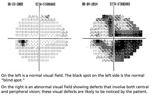
- Automated visual field test – this tests the sensitivity of the peripheral vision, where glaucoma strikes first and is performed using the Humphrey Visual Field Analyser, a computer-controlled test in a darkened room.
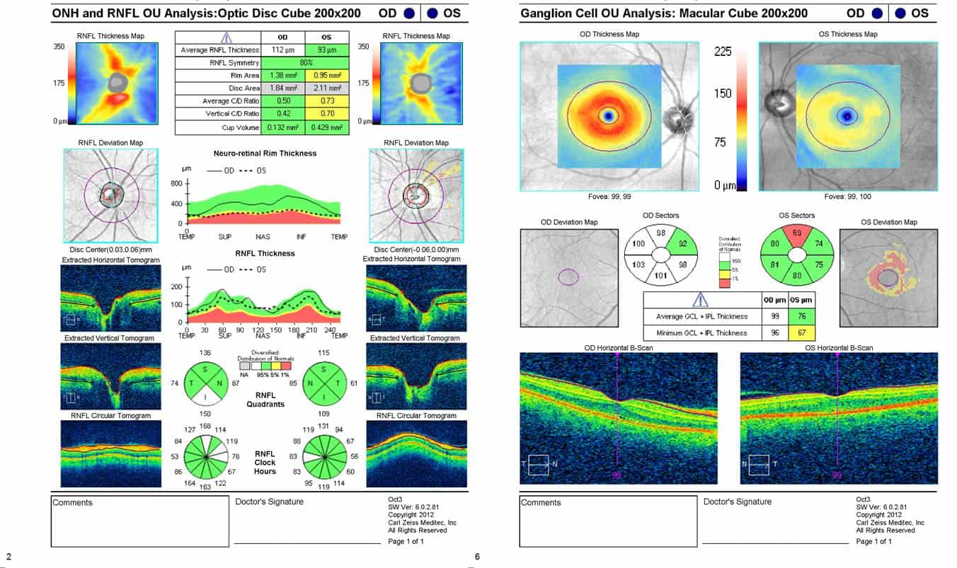
- Measurement of the optic nerve and retinal nerve fibre layer by HRT or OCT.
- Examination of your optic nerve and review of test results by your Ophthalmologist.
- Performed every 6-12 months or as requested by Doctor
- Tests peripheral vision
- The Optic nerve becomes compressed due to increased pressure in the eye which causes loss of peripheral vision
- This test can be done at our rooms in Murdoch or Subiaco
- Time – takes approximately 20 mins
The Heidelberg Retina Tomograph is a confocal laser scanning system for imaging of the front and back of the eye. It comprises various modules which can be used independently or combined with each other. The major routine clinical application is the analysis of the optic nerve head structure for glaucoma diagnosis. A second application is in diabetes for the location and quantification of retinal edema.
The HRT system is tried-and-tested, versatile technology.
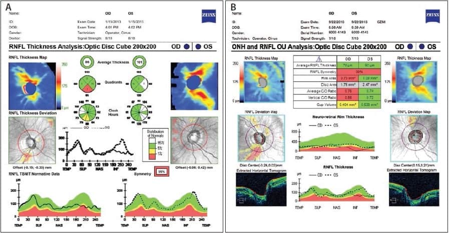
Optical coherence tomography (OCT) is an imaging technique that uses coherent light to capture to micron-level resolution, 2- and 3-dimensional images from within the eye.
Ocular (or ophthalmic) OCT is used heavily by ophthalmologists to obtain high-resolution images of the retina and anterior segment of the eye. Owing to OCT’s capability to show cross-sections of tissue layers with micrometre resolution, OCT provides a straightforward method of assessing the cellular organization, photoreceptor integrity, and nerve fibre layer thickness in glaucoma, macular degeneration, diabetic macular edema, multiple sclerosis and other eye diseases or systemic pathologies which have ocular signs.